The femur is the longest and most powerful bone in the body. It lies between the hip and knee. To break this bone, a lot of force is needed. A femur fracture is a critical example of bone fracture and needs immediate medical attention.
Rehabilitation, including physical therapy, is essential for regaining proper function of different parts of the body and keeping it mobile. Such fractures are injuries that need urgent medical attention. This is done to avoid late complications and boost an early recovery. It takes weeks and sometimes months to fully recover from this fracture.
What Makes this Fracture a Serious Injury?
Reasons that make this fracture serious:
- Loss of blood, as there is a good amount of internal bleeding.
- As it involves a sizable pain, one may get a shock.
Causes of Femur Fracture
Aged people over 65 years of age have an increased tendency to break their femurs if proper care is not taken. The most common causes of such bone fracture are:
- Motor Vehicle Accidents: These are more prominent in young individuals.
- Falls are a Significant Cause of such Fractures: These may show up in children and elderly persons with fragile bones.
- Child Maltreatment: Newborns and young children are more prone to this fracture.
- Gunshot wounds are a prevalent cause of such fractures.
- Sports injuries are also common in young and active sports people.
Symptoms of Femur Fracture
If you’ve fallen or injured, you might have a broken femur, and the given signs may be of concern:
- An intense and unbearable pain is causing you restlessness.
- You are not able to put weight on your wounded leg.
- You’re bleeding because pieces of your femur have broken through the skin. This is known as an open fracture.
- Pieces of your femur push up against your skin without piercing it. This is known as a closed fracture.
- Your thigh is bruised.
- Parts of your thigh are enlarged.
- Your wounded limb may be shorter than your intact leg, and it may turn away from your body.
Diagnosis and Tests of Femur Fracture
A healthcare practitioner will thoroughly examine the damaged leg for abnormalities, edema, and discomfort. The patient’s capacity to support weight and perform specific activities is also assessed. A femur fracture is diagnosed by scrutiny of the damaged leg. The healthcare will obtain your X-rays.
- X-rays: It is one of the main diagnostic tools. It determines the damage done to the femoral fracture. These provide detailed images that aid in the diagnosis of fractures and treatment options.
- CT Scans: When more details of the fracture is required, computed tomography (CT) scans are helpful. It may be used to generate detailed cross-sectional images of the fractured femur.
- MRI (Magnetic Resonance Imaging): It can be used to evaluate soft tissue injuries, such as ligaments.
Treatment for Femur Fractures
The treatment for femur fractures mainly revolves around the specific nature of the fracture.
Non-Surgical Method
- Casting: Fractures particularly in youngsters or less severe instances make use of this technique. It is used to immobilize the leg and aid recovery. This can be made of plaster or fiberglass.
- Traction: Regular pulling force is applied to the affected leg. It makes use of weights and pulleys. The objective in this technique is to straighten and stabilize bones, especially in emergency settings.
- External fixation: Metal pins or screws are inserted into the bone that lies above and below the fracture site. It is then joined by an external frame. This makes the fracture stable. And gradually enables for healing without requiring direct surgical intervention.
Surgical Method
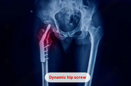
- Intramedullary Nailing: It is a common surgery and includes putting a metal rod (nail) into the femur’s hollow interior to fix the fracture. This strategy allows for early movement and decreases the likelihood of non-unionization.
- Open Reduction and Internal Fixation (ORIF): This medical procedure involves stabilizing and realigning of fractured bones. Later these are secured using screws, plates, or other internal devices. ORIF is commonly used for complicated or displaced fractures.
- Plate Fixation: Plates and screws are made use in this procedure. It stabilizes the broken pieces of bones. Where intramedullary nailing is not possible, this approach is considered.
Risks Associated with Broken Femur Surgery
As with any surgery, this fracture too has some potential perils, and these include:
- The broken femur bone may not join correctly. This may call for a longer healing period or more X-rays will be required to see where the problem is.
- The nail and screws put to hold the broken bones may cause irritation.
- Clotting of blood may also take place.
Taking Care After the Surgery
Try sleeping flat on your back. Keep the broken femur high above your heart level. In this manner, this position will keep your bone away from swelling. Take the following actions that will decrease the danger of falling:
- Tripping hazards, such as loose cords and throw rugs should be avoided.
- Make sure to install rails on both sides of the staircase.
- Keep frequently used goods in cabinets. It will make easy access without the use of a step stool.
- Use non-slip mats in the bathtub and on the shower floor.
For full recovery it may take four to six months and it depends on factors like age, how deep is the fracture. By performing exercises that focus on improving the range of motion of joints, you will be able to recover fast.