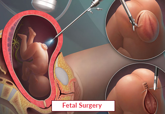
Spina bifida affects pregnant women. It happens due to inappropriate formation of the spine and spinal cord in the womb. This neural tube defect leads to life-threatening conditions such as chiari malformation, orthopedic problems, latex allergy etc., if it is left untreated. It is a rare or severe health issue, which cannot be cured easily. Its most common approach is fetal surgery. This procedure is applicable for the improvement of outcomes and can be performed though fetoscopic surgery, open surgery and other surgical techniques. It is also known as in-utero or prenatal surgery.
What is a Neural Tube?
A neural tube is a tube-shaped structure, which develops in the central nervous system. It forms and closes in the embryo during pregnancy for third and fourth weeks. Its upper part appears in the brain and skull, whereas the lower part originates in the spinal cord and the backbone.
When it fails to close completely during pregnancy, then it causes birth defects. It leads to spina bifida and can be harmful for one’s health. A pregnancy problem can occur in women and they face trouble while giving birth to their children.
Why does the Fetal Surgery Procedure Need to be Performed?
This surgical procedure is applicable for the treatment of different health conditions. These are as follows:
- Fetal anemia
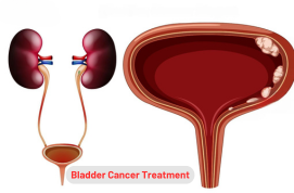
- Bladder outlet obstruction
- Mediastinal teratoma
- Amniotic band syndrome
- Partially or fully blockage of airway in fetus
- Lump or swelling in neck
- Growth of tumor in baby’s tailbone
- Pulmonary Sequestration in lungs
- Monochorionic twin complications
It is not recommended for patients with chromosomal abnormalities, maternal HIV etc. They must consult with their doctor for these health issues.
Advantages of Fetal Surgery Treatment
This procedure has several benefits. These include the following:
- This approach is recommended for fetus protection.
- It treats birth defects to improve a long-term outcome of an unborn baby and maintains a better-quality life.
- A few surgical procedures are needed for spina bifida treatment. The fluid gets diverted away from the brain by the first birth of a child.
- The motor skills can become better in children with two and a half years of age while walking without crutches or other devices.
Risks and Complications of Fetal Surgery
This surgery is safe and effective for patients but it leads to several complications. These are as follows:
- Loss of blood
- Thinning or re-opening in the uterine scar
- Growth of gestational diabetes
- Infection
- Premature labor
- Fatal death
- Infertility
- Lower blood pressure or breathing problems from medication drugs.
Diagnosis for Fetal Surgery Treatment
This procedure uses two essential ways to diagnose fetal issues in patients. These are as follows:
- Blood Sample: A small amount of blood is to be removed from the fetus through a very small needle. It is mainly recommended for the identification of a type of blood, fetal anemia, and low platelet count. A surgeon performs this procedure for the diagnosis of fetal infections, chromosome abnormalities etc.
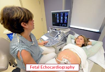
- Fetal Echocardiography: The heart of an unborn baby can be checked with its position, size, function, structure and rhythm. It is mainly suitable to diagnose congenital heart disease in patients. This procedure is helpful for early genetic testing and can be performed between 11 and 14 weeks of pregnancy.
Treatment Options for Fetal Surgery
The treatment approaches of this surgery are as follows:
Fetoscopic Surgery
A small incision is made in the uterus and the tiny camera known as a fetoscope is attached on the end of a long, fiber-optic tube. After that, they use long-narrow tools for the operation. This method is minimally invasive and can be suitable for the management of twin-to-twin transfusion syndrome or diaphragmatic hernia.
The surgeon opens the abdomen and makes two or three incisions to place the camera and instruments inside the uterus. This process is helpful for the treatment of myelomeningocele in patients.
Open Surgery
A general anesthesia will be provided to women and then an incision can be made in an abdomen and uterus to reach the fetus. After reaching, a surgeon performs a surgical operation inside it. The uterus and belly are then closed and the pregnancy will continue to be as normal as possible.
Fetoscopic EndoTracheal Occlusion
A congenital diaphragmatic hernia can be managed when it becomes an unborn baby. This condition occurs due to development of holes in a diaphragm. The small instrument known as fetoscope is attached with the uterus for fetus and placenta visualization. A trachea with a latex balloon can be blocked for the promotion of lung development of a baby.
Shunt Placement
A surgeon can drain the fluid from the amniotic cavity by placing a hollow tube, known as shunt, to the fetus. It is an outpatient procedure, which treats certain health issues such as blockage in urinary tract, excess fluid in the chest etc.
Intrauterine Transfusion
A surgeon applies an injection on a red blood cell for the management of severe fetal anemia. It is a successful treatment, which can be performed through intravascular or intraperitoneal transfusion methods.
Ex-utero Intrapartum Treatment
The oxygen and blood are received through the placenta before childbirth. This procedure is recommended for babies with airway compression, neck or face tumors, micrognathia, etc. It is safe and effective for them.
Our Other Services
Latest Health Tips
Can Immunotherapy Cure Stage 4 Lung Cancer?
Early Signs of Cervical Cancer
Foods that Kill Cancer: Leafy Vegetables, Grains, & More
What Stage of Cancer is Immunotherapy Used For?
Which is Worse for Cancer, Sugar or Alcohol?
Vaccines That Prevent Cancer
What Kills Cancer Cells in the Body Naturally?
Early Warning Signs of Bone Cancer
Submit Your Enquiry
Testimonials


